Two Early BBRF Grants Helped Launch the Career of the Nation’s New NIMH Director
Joshua Gordon, M.D., Ph.D., remembers being deeply impressed during college with a mentor who was devoted not just to research—about genes whose activation sends cells on a path toward cancer`— but also to using knowledge about these “oncogenes” to help doctors all over the world assess their patients’ tumors, enabling better treatment decisions.
“That marriage between learning something basic about how biology works and intimately linking it with issues of direct relevance to patient care” not only impressed Gordon, but became a model for his future work in neuroscience.
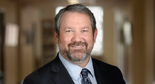
In July 2016, the Director of the National Institutes of Health, Dr. Francis Collins, announced that Dr. Gordon, most recently of Columbia University, would become the next Director of the National Institute of Mental Health (NIMH), filling a vacancy left by the departure of Dr. Thomas Insel, who led the Institute for the past 13 years.
Dr. Gordon, who received career-shaping BBRF Young Investigator Grants in 2001 and 2003, was described by Dr. Collins as “a visionary psychiatrist and neuroscientist with deep experience in mental health research and practice.” He is the fifth individual with important connections to the Foundation to lead our nation’s agency devoted to advancing research and treatment of mental illness—the largest such institution in the world.
Past leaders of the NIMH include Herbert Pardes, M.D., Founder and President of the Brain & Behavior Research Foundation’s Scientific Council, and Foundation Scientific Council Members Frederick Goodwin, M.D., Stephen Hyman, M.D., and Lewis Judd, M.D.
We spoke with Dr. Gordon about his goals for the NIMH and about his own research on brain circuitry whose promise helped catapult him into a position of international prominence in neuroscience. One of Dr. Gordon’s foremost priorities at the NIMH echoes the lesson of his college mentor. “I have an interest in trying to move from techniques that enable us to learn about the brain—by studying how its neural circuits work—toward ways of using this new knowledge to develop new treatments.” His own lab’s recent research demonstrates how the two paths —basic and applied research—can support and amplify one another.
In both mental health and mental illness, the importance of neural circuits—whose trillions of connections give rise to consciousness, enable perception, and make possible all of our responses to the world around us—is self-evident. But how to make sense of these myriad linkages over which messages between hundreds of millions of neurons pass every second and how to mobilize this knowledge in developing next-generation treatments—these are among the great questions of modern science.
Dr. Gordon’s devotion to the brain and to a career in neuroscience began with neural circuits. In 1990, he was attending a conference in Washington, D.C. and heard a lecture about micro-stimulation. “The investigators used small electric currents to change the activity patterns in a monkey’s brain. As the patterns changed, so did the monkey’s behavior. That was astonishing to me. It was a fascinating, if rudimentary, example of the relationship between patterns of brain activity and patterns of behavior,” Dr. Gordon remembers.
Immediately upon returning to San Francisco, where he was then in the early part of his medical school training at U.C. San Francisco, Gordon told those in charge that he was switching his focus from molecular biology to neuroscience.
When he applied for his first BBRF Young Investigator Grant in 2000, Dr. Gordon was in the fourth and final year of his medical residency and performing research in the laboratory of René Hen, Ph.D., at Columbia University (a 2009 and 2003 Distinguished Investigator, a 1998 Independent Investigator, and a member of the Scientific Council). Gordon’s project involved recording patterns of brain activity during behavior, which called for equipment the Hen lab did not have.
Nobel prizewinner Dr. Eric Kandel (also of the Scientific Council) graciously offered Gordon access to equipment in his own lab, but Gordon’s research time was limited. “I applied for a ‘NARSAD,’ and when I received that first grant I went out and bought myself a rig. It was the fact that I could go to BBRF and get this early support that enabled me to launch my career,” Dr. Gordon says.
His second BBRF Young Investigator Grant was awarded in 2003, when Gordon was a research fellow at Columbia, “and it completely enabled me to jump start things when I joined the faculty in 2004.” At Columbia, as elsewhere in the U.S., a new assistant professor is obliged to secure research funding, usually from the government, to sustain a laboratory over a period of years. “Without that second Young Investigator Grant I honestly wouldn’t have had nearly enough time on the equipment I needed to produce the data that enabled me to get that essential government award and make the transition to research independence. Dr. Gordon was, in other words, the perfect example of the sort of researcher the Young Investigator Grants seek to identify and help: one who is unlikely to gain independence until s/he can generate experimental results of sufficient promise to draw a much larger and sustaining government grant.
Partly because of the way grants are reviewed at the government level—the need for positive early results before multiyear support is extended—one appreciates the importance of the Brain & Behavior Research Foundation and other philanthropies that support promising but as yet unproven young people beginning a career of research, whether in neuroscience or clinical psychiatry.
“Just looking at my case it’s pretty clear there are gaps, where government funding isn’t there,” Dr. Gordon says. “And while it’s reasonable to ask how we might fill those gaps from a government perspective, the fact that a philanthropic organization like the BBRF can jump in, in a much more flexible way, is incredibly crucial to the research enterprise.”
IN SCHIZOPHRENIA, THE PROSPECT OF RESTORING NEURAL SYNCHRONY TO ADDRESS COGNITIVE DEFICITS
Long before the advent of modern neuroscience, those who have cared for and spent time with people living with schizophrenia have described the disorder in terms of a “disconnection.” Patients who experience the hallucinations and delusions that characterize psychosis, for example, may seem to be “disconnected” from the reality that others around them are experiencing.
The research of Dr. Joshua Gordon and his colleagues has provided scientific support for the notion of schizophrenia as a kind of “disconnection syndrome,” although in a way that is not at all obvious, and in fact doesn’t refer to dramatic, manifested symptoms like a person hearing voices. Rather, Dr. Gordon’s work has shed new light on the so-called cognitive symptoms of schizophrenia: debilitating yet less visible deficits in memory, attention and learning. These are aspects of the illness that tend to, more often than other symptoms, prevent people from integrating successfully in society. That’s because they directly affect their capacity to think clearly, communicate, and work.
Dr. Gordon’s research has centered on the concept of neural synchrony. “Synchrony,” he explains, “literally means things that are happening together in time.” In the brain, synchrony can be measured in two basic ways. “At the local level — say, in a single area of the brain like the hippocampus or the cortex, synchrony can mean that neurons within that area tend to fire together, and become silent together.” In the language of neuroscience, neurons within brain regions can exhibit synchrony in activation and inhibition.
The other fundamental kind of synchrony in the brain is coordination across different brain regions. “This makes intuitive sense,” Dr. Gordon suggests. “Imagine, for instance, that the hippocampus needs to work together with the prefrontal cortex on a particular task. To accomplish this work, you would imagine that things work better when the two regions are working in synchrony.”
While a logical idea, in science, this is really nothing more than a hypothesis. It needs to be proven in order for us to learn anything useful from it. This is exactly what some of Dr. Gordon’s research has accomplished. It turns out that different kinds of synchrony have been observed in the brain using different technologies. Watching parts of the brain function in an MRI scanner while a person performs a task has, for instance, revealed certain patterns of activity within and between brain regions. Those patterns play out on a time scale of seconds.
The kind of patterns Dr. Gordon scrutinizes occurs over much more compressed time intervals, on the order of hundredths of a second. His focus is on patterns that arise from electrical activity in the brain—activity that helps move messages from neuron to neuron in the almost incomprehensibly dense thicket of cells and the “wires” that are packed into our most complex organ. Electrical patterns—popularly referred to as brain waves—have long been measurable with a technology called EEG—electroencephalography.
With EEG, characteristic patterns of fluctuation in electrical activity within and across brain regions have been delineated. Within a structure like the hippocampus, for example, one fluctuation in electrical signals, called a theta wave, warbles Joshua Gordon, M.D., Ph.D.
eight times per second in what Dr. Gordon describes as a kind of “up—and—down, up—and—down” pattern. At the same time, other types of waves “ride” on top of that signal. Gamma waves in the hippocampus fluctuate 40 times per second. When the hippocampus is in synch, the relation of the slower wave to the faster is such that together they appear almost to regulate one another—the frequency of each keying off of and thus appearing to keep that of the other “in synch.”
These patterns are more than mere scientific curiosities. In 2010, Dr. Gordon, working with his frequent collaborator Dr. Joseph A. Gogos of Columbia University, an authority on the genetics of schizophrenia who has developed and studied several important mouse models of the illness, studied brain waves in mice as they tried to navigate through a maze. Their experiments showed that there is synchrony not only locally, within two key brain regions—the hippocampus and the medial prefrontal cortex—but between them as well.
“Not only can we measure that there is synchrony, but we can measure how strong it is,” Dr. Gordon explains. “It depends on what the animal is doing, behaviorally.” In making its way through a maze, a rodent has to process information about where it is—spatial information—and it must remember where it has just been— tapping short-term memory—if it is to reach the piece of cheese at the end of the maze. In a healthy mouse, “when the animal is doing a working memory task [like remembering where it has just been], you see an enhancement in synchrony between the hippocampus and the prefrontal cortex,” says Dr. Gordon. But here’s the key point: things were different in the mice that Dr. Gogos had developed to mimic some of the key symptoms of schizophrenia.
These mice were bred with a genetic defect called 22q11 deletion syndrome—a mutation on chromosome 22 that is seen in a subset of people with schizophrenia and is thought to be powerful enough to cause the illness. When these mice navigated a maze, synchrony between the hippocampus and prefrontal cortex was weaker than in healthy mice. “This suggested to us that perhaps working memory is impaired in schizophrenia because those two brain regions are not working together properly,” Dr. Gordon says.
In the “schizophrenia” mice, the two regions were not dis-connected so much as dys-connected; they did not seem to be working in optimal synchrony. Interestingly, these experiments revealed that there was no problem with synchrony among neurons within each of the two regions, only between them. This led to more experiments, culminating in a 2015 paper in which Dr. Gordon and colleagues intentionally disrupted long-range connections between the two regions in healthy mice, and observed impairments in the animals’ working memory capacity. Disrupting specific kinds of brain waves led to another interesting observation: what is specifically lost in the observed dys-synchonry between the prefrontal cortex and hippocampus was the mouse’s ability to encode new information and thus keep in mind the spatial location of its goal as it navigated a maze.
In 2016, another exciting step forward in the research: in mice with 22q11 deletion syndrome, modeling human schizophrenia, Dr. Gordon and colleagues confirmed that working memory deficits precisely affected the animals’ ability to mentally represent the location of their goal, due to a lack of synchrony between the hippocampus and prefrontal cortex. What remains to be shown, Dr. Gordon says, is whether the problem identified in the 22q11 mice crops up in mice with other genetic defects that give rise to schizophrenia-like symptoms. Another question, more difficult to answer, is whether people with 22q11 deletion syndrome have the synchrony problem that mice do.
The most exciting question, if the synchrony problem is confirmed in people, is whether it can be reversed. Dr. Gordon thinks there are several ways to attempt this, and with Dr. Gogos and colleagues has already tested one in mice, with results he calls “flabbergasting.” In mice with 22q11 deletion syndrome, the scientists gave a drug during the fetal development period. The drug inhibits GSK3, one of many proteins disrupted because of the 22q11 mutation. Administering this drug appeared to restore the integrity of long-range connections from the prefrontal cortex to the hippocampus and boosted working memory in the mice, after birth. “Whether this will be applicable in people I don’t know,” Dr. Gordon says, “but it is an astounding and wonderfully promising result.”
In a recent article in the journal Science reviewing advances made by another neuroscience team studying the brain, Dr. Gordon likened a brain affected by a serious neurodevelopmental disorder to “a tangled mess of threads.” The point of the metaphor, he noted in our conversation, is not to stress the tangle, but rather the absolute necessity of “following the threads all the way through,” wherever they lead. “That’s how we learn something about these devastating illnesses. We have to have the patience to learn how to untangle what is tangled—something I vividly remember my mother helping me to do when I was very young."
— Written By Peter Tarr
Click here to read the Brain & Behavior Magazine's March 2017 issue